Home

socks judge gone crazy bregma mouse brain jump curb Pinpoint

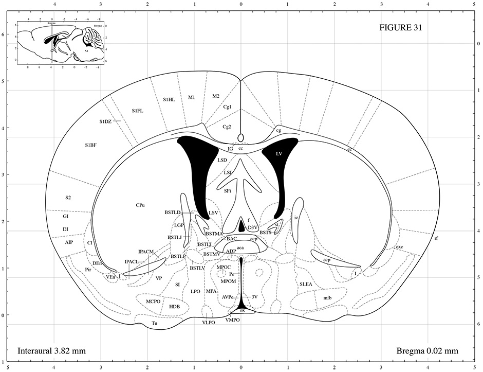
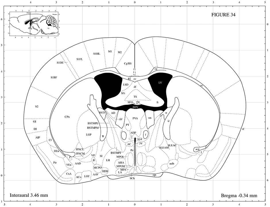
Mouse Brain Atlas
Mouse Brain Atlas

Metabolic Mapping of Mouse Brain Activity after Extinction of a Conditioned  Emotional Response | Journal of Neuroscience
Metabolic Mapping of Mouse Brain Activity after Extinction of a Conditioned Emotional Response | Journal of Neuroscience
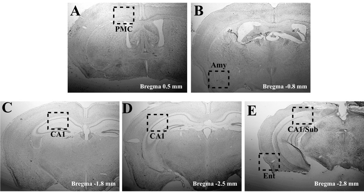
Detailed immunohistochemical characterization of temporal and spatial  progression of Alzheimer's disease-related pathologies in male  triple-transgenic mice | BMC Neuroscience | Full Text
Detailed immunohistochemical characterization of temporal and spatial progression of Alzheimer's disease-related pathologies in male triple-transgenic mice | BMC Neuroscience | Full Text

Mouse Brain Atlas
Mouse Brain Atlas

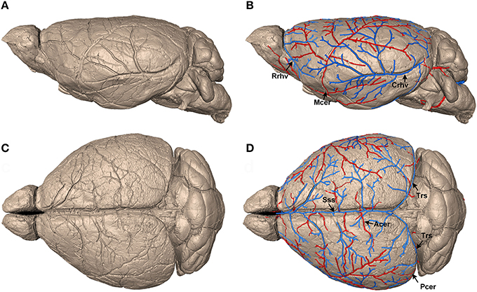
Frontiers | Precise Cerebral Vascular Atlas in Stereotaxic Coordinates of  Whole Mouse Brain
Frontiers | Precise Cerebral Vascular Atlas in Stereotaxic Coordinates of Whole Mouse Brain

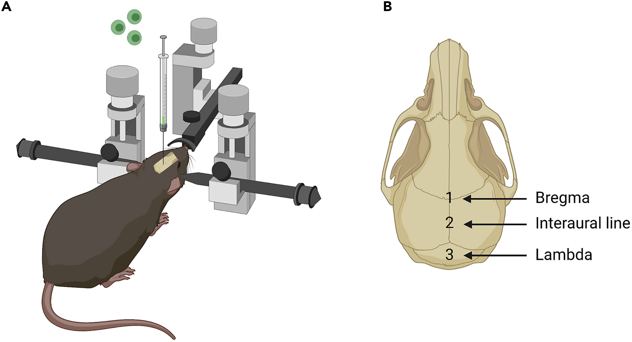
An optimized intracerebroventricular injection of CD4+ T cells into mice

Mouse EEG - channel and source analysis - FieldTrip toolbox

GABA and Glutamate Pathways Are Spatially and Developmentally Affected in  the Brain of Mecp2-Deficient Mice | PLOS ONE
GABA and Glutamate Pathways Are Spatially and Developmentally Affected in the Brain of Mecp2-Deficient Mice | PLOS ONE
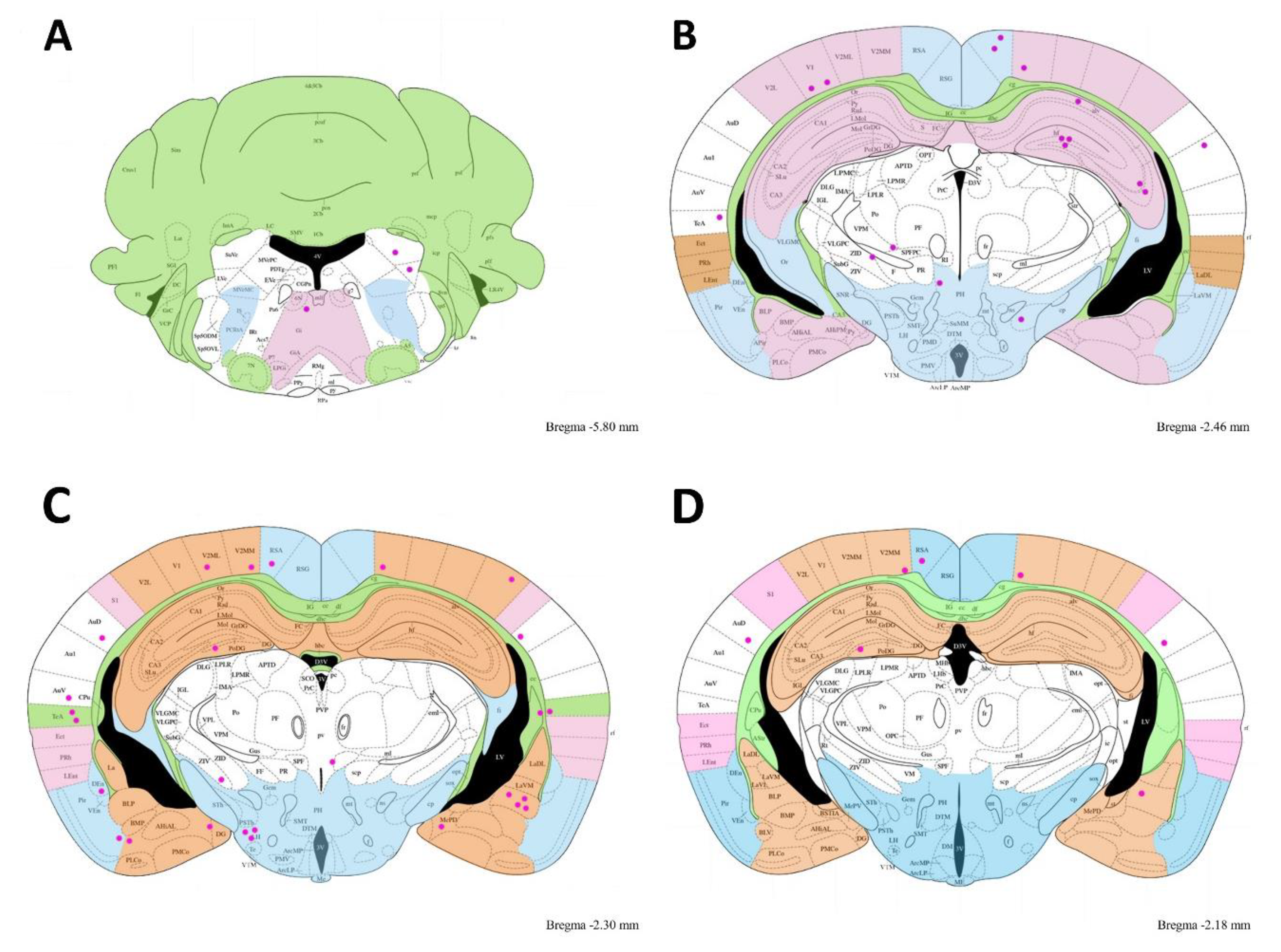
Microorganisms | Free Full-Text | Kinematic Locomotion Changes in C57BL/6  Mice Infected with Toxoplasma Strain ME49
Microorganisms | Free Full-Text | Kinematic Locomotion Changes in C57BL/6 Mice Infected with Toxoplasma Strain ME49

Mouse Brain Atlas
Mouse Brain Atlas

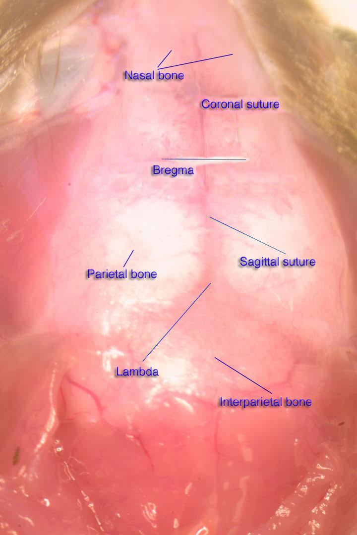
Where do you measure the Bregma for rodent stereotaxic surgery? -  ScienceDirect
Where do you measure the Bregma for rodent stereotaxic surgery? - ScienceDirect

Mouse Brain Gross Anatomy Atlas

Procedure to determine the stereotaxic coordinates (A) Illustration... |  Download Scientific Diagram
Procedure to determine the stereotaxic coordinates (A) Illustration... | Download Scientific Diagram

Figure 6 from Phosphorylation of Histone H2AX in the Mouse Brain from  Development to Senescence | Semantic Scholar
Figure 6 from Phosphorylation of Histone H2AX in the Mouse Brain from Development to Senescence | Semantic Scholar
Mouse Brain Atlas
Mouse Brain Atlas

Frontiers | Stereotaxic atlas of the infant rat brain at postnatal days 7–13

Delivery of Antisense Oligonucleotides to the Mouse Brain by  Intracerebroventricular Injections | SpringerLink
Delivery of Antisense Oligonucleotides to the Mouse Brain by Intracerebroventricular Injections | SpringerLink
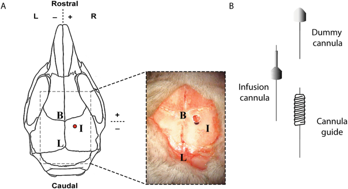
3. The position of bregma on the mouse skull. (A) The mouse skull,... |  Download Scientific Diagram
3. The position of bregma on the mouse skull. (A) The mouse skull,... | Download Scientific Diagram

Multiplex Three-Dimensional Brain Gene Expression Mapping in a Mouse Model  of Parkinson's Disease
Multiplex Three-Dimensional Brain Gene Expression Mapping in a Mouse Model of Parkinson's Disease

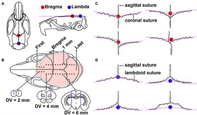
Semi-automatic extraction of bregma and lambda from micro-CT skull... |  Download Scientific Diagram
Semi-automatic extraction of bregma and lambda from micro-CT skull... | Download Scientific Diagram

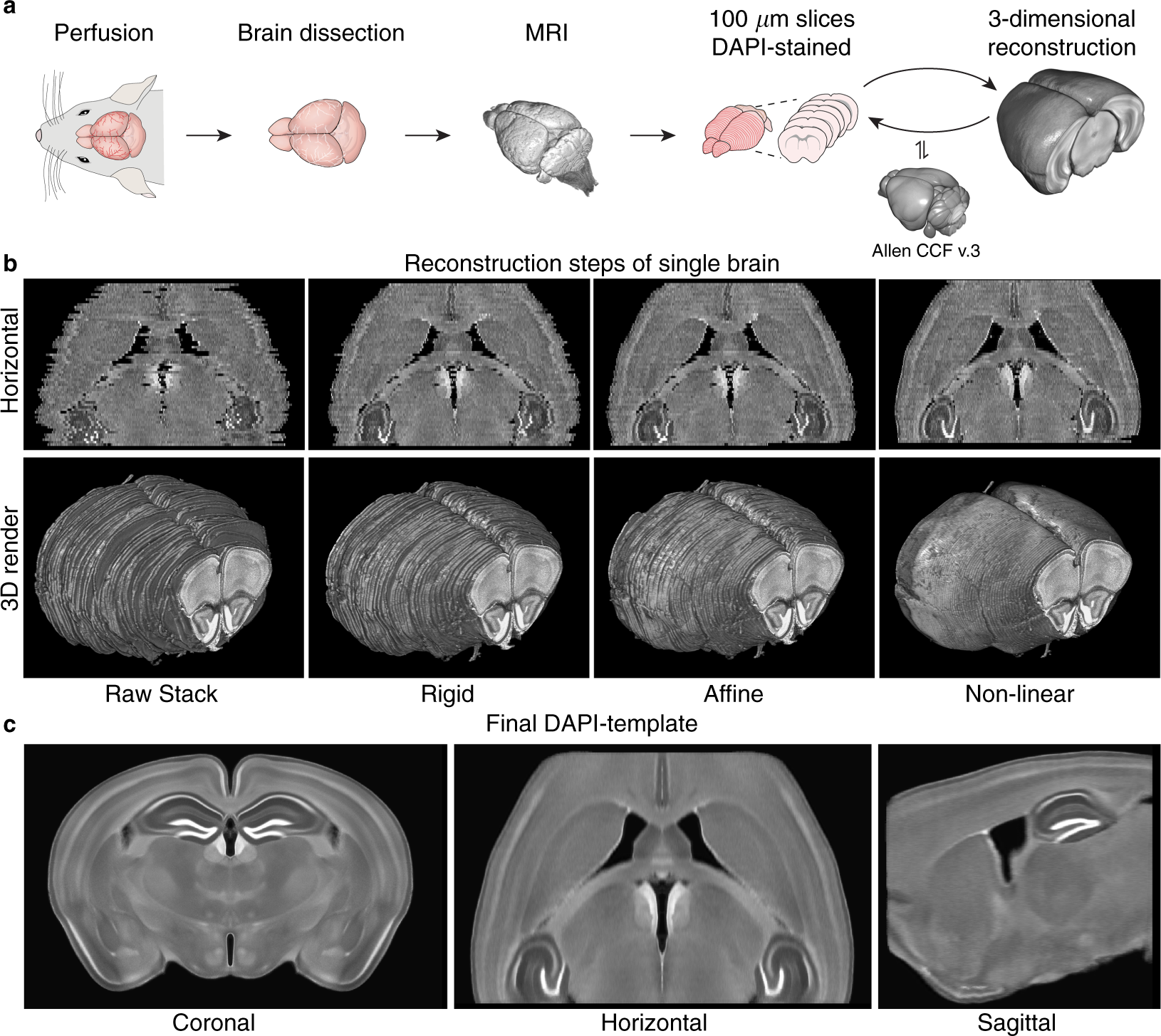
A three-dimensional, population-based average of the C57BL/6 mouse brain  from DAPI-stained coronal slices | Scientific Data
A three-dimensional, population-based average of the C57BL/6 mouse brain from DAPI-stained coronal slices | Scientific Data

Multimodal 3D Mouse Brain Atlas Framework with the Skull-Derived Coordinate  System | Neuroinformatics
Multimodal 3D Mouse Brain Atlas Framework with the Skull-Derived Coordinate System | Neuroinformatics

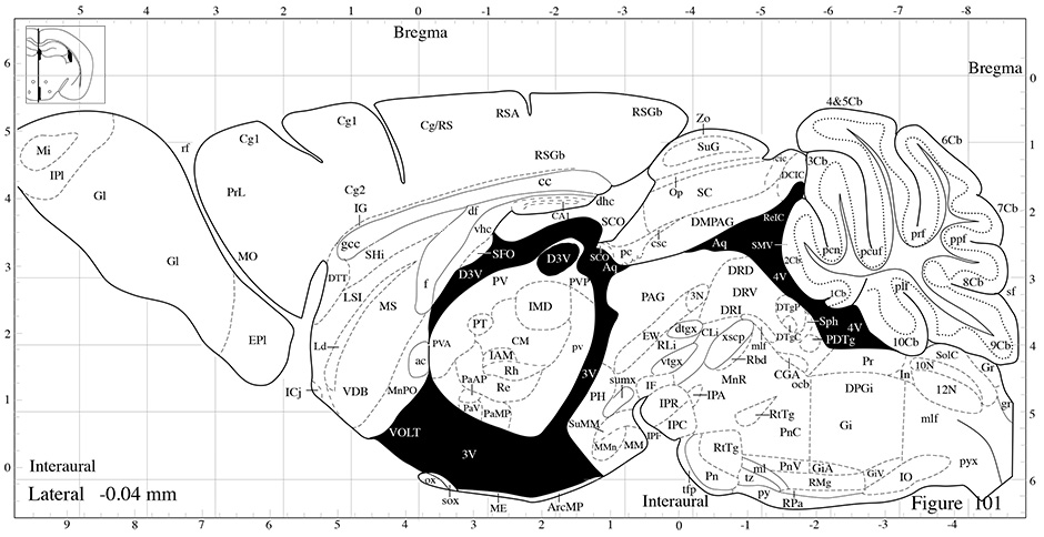
Drawings taken from the mouse brain atlas of Paxinos and Franklin [40]  showing the location of forebrain regions where Fos-ir cells were  quantified (shaded areas in each panel).
Drawings taken from the mouse brain atlas of Paxinos and Franklin [40] showing the location of forebrain regions where Fos-ir cells were quantified (shaded areas in each panel).

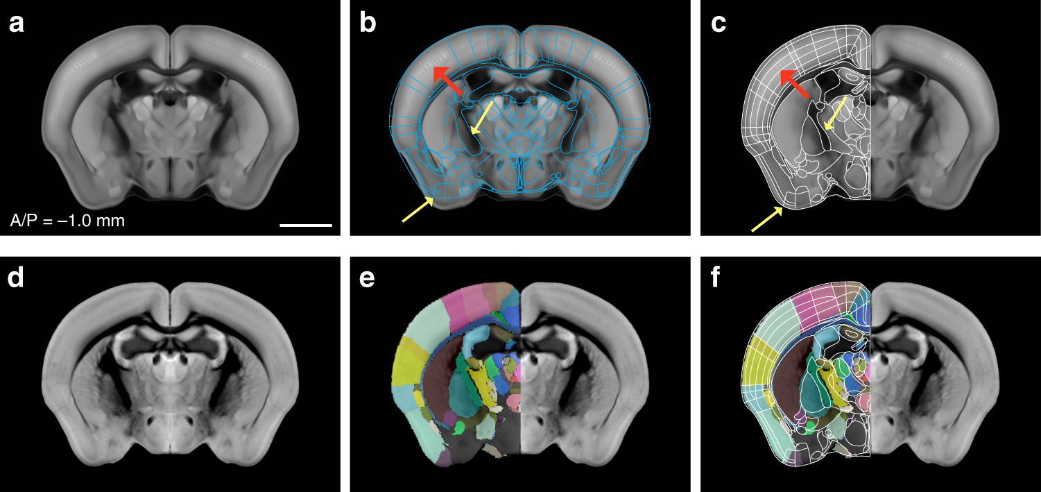
Enhanced and unified anatomical labeling for a common mouse brain atlas |  Nature Communications
Enhanced and unified anatomical labeling for a common mouse brain atlas | Nature Communications

What is the typical distance from Bregma to Lambda in adult mouse brains? |  ResearchGate
What is the typical distance from Bregma to Lambda in adult mouse brains? | ResearchGate

Striatal Infusion of Glial Conditioned Medium Diminishes Huntingtin  Pathology in R6/1 Mice | PLOS ONE
Striatal Infusion of Glial Conditioned Medium Diminishes Huntingtin Pathology in R6/1 Mice | PLOS ONE